Focus area
Dual-energy & Spectral CT
We quantitatively examine material decomposition, virtual monoenergetic imaging, and beam-hardening effects to improve the reliability of basis-material density images.
Page views Today — · Total —
 Kondo CT Imaging LabKyushu University
日本語
Kondo CT Imaging LabKyushu University
日本語
Science of CT Imaging · Fukuoka, Japan
We make the most of existing medical equipment and imaging data to advance greener radiology.
Research
Focusing on dual-energy CT, low-dose CT, and image reconstruction, we revisit CT imaging from the perspective of radiation physics and work to improve the accuracy and reproducibility of quantitative image evaluation.
Focus area
We quantitatively examine material decomposition, virtual monoenergetic imaging, and beam-hardening effects to improve the reliability of basis-material density images.
Focus area
We investigate the relationships among image noise, reproducibility, and acquisition parameters to preserve diagnostically useful information while reducing radiation exposure.
Focus area
We analyze how reconstruction algorithms and scan geometry affect CT images and develop predictable, reproducible methods for image evaluation.
Our approach
Starting with system physics, we ask why CT images look the way they do, measure the underlying effects, test reproducibility, and translate the findings into clinically useful methods.
Measure
We describe image quality and errors that are difficult to judge visually in terms of physical and statistical quantities.
Validate
Through phantom experiments and repeated measurements, we test whether findings remain valid across systems and acquisition conditions.
Translate
We translate insights from imaging science into methods that can improve diagnostic accuracy and reduce patient burden.
Free research web tool
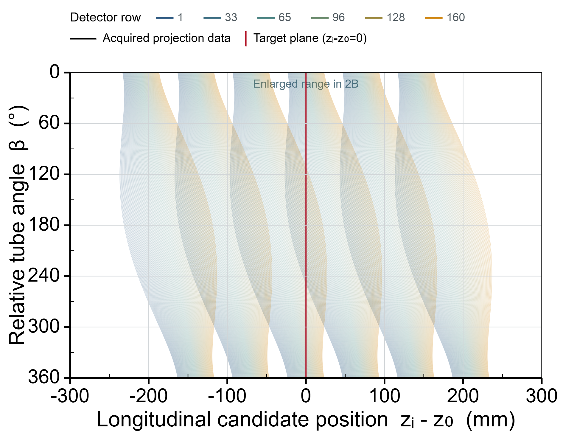
Free to use in your browser; no registration or installation is required. Explore how detector-row geometry, beam pitch, transaxial position, and configured slice thickness affect longitudinal candidate-ray positions, linear interpolation weights, and an explanatory longitudinal slice sensitivity profile (SSPz). The model uses acquired projection data over a 0–360° full scan and does not emulate a proprietary commercial reconstruction.
Open the free SSPz geometry referenceUseful —
Free web tool
Optimized for smartphones and tablets. Set presentation and discussion times in minutes and seconds, then use the large countdown, selectable alert sound, and one-tap next-speaker restart.
Open the timerUseful —

Free web tool
Set lesson start and end times and play a school chime according to network-synchronized time. Designed for iPad and browser use, with editable schedules, weekday selection, volume control, and Japanese and English interfaces.
Open the school bell toolUseful —

Highlights
Our latest research presentation and professional recognition. A complete chronological record is available in the achievement archive.
Joint 5th Place · Top 12 Presentations
Energy Dependence of Water-Density Correction in Virtual Monoenergetic Images of Dual-Energy CT
Koki NanjoGraduate School of Medical Sciences, Kyushu University
82nd Annual Meeting of the Japanese Society of Radiological Technology
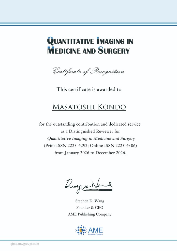
View official announcementDistinguished Reviewer
Recognized for outstanding contribution and dedicated service as a Distinguished Reviewer.
Masatoshi KondoKondo CT Imaging Laboratory
 View certificate ↗
Podcast
Short audio briefings on radiology, medical imaging, medical AI, conferences, and recent research—available on Spotify and Apple Podcasts.
 日本語
日本語
Japanese edition
Latest episodeEpisode #15
Spotify Apple Podcasts English
English edition
Latest episodeEpisode #11
Spotify Apple Podcasts 한국어
한국어
Korean edition
Latest episodeEpisode #12
Spotify ภาษาไทย
Thai edition
Latest episodeEpisode #12
SpotifyCollaboration
We welcome research discussions with medical institutions, universities, and industry, as well as enquiries about graduate research. Please feel free to contact us even if the connection with our current themes is still taking shape.
Profile
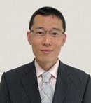
Division of Medical Quantum Science, Department of Health Sciences, Faculty of Medical Sciences, Kyushu University
